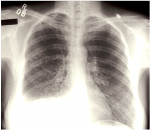
Case 1. CXR of patient (a) with right pleural effusion and cystic lung disease
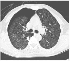
Case 1. CT Scan of the lungs (b) shows typical cysts found in patients with Lymphangioleiomyomatosis (LAM)
38 year old female noted right sided chest pain for 3 days. She had a background of difficulty in breathing over the past 6 months. When she was seen, she was found to have a right pleural effusion (fluid on the skin of the right lung) with pneumothorax (air trapped between the right lung and chest wall). Thoracocentesis (removing the fluid with a needle) showed that the fluid was milky white, typical of chylothorax. Review of the Chest Xray (a) also showed that the lung fields were abnormal. CT Thorax (b) showed diffuse lung cysts, typical of a rare condition called LAM.
This disease is relatively rare and almost exclusively affects females, typically in their child bearing age. This disease is characterised by abnormal growth of smooth muscle cells in the lungs, lymphatics and kidneys.
https://www.thelamfoundation.org/Newly-Diagnosed/Learning-About-Lam/About-LAM
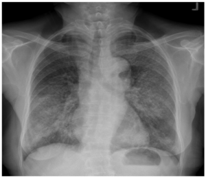
Case 2- CXR (a) of patient showing bilateral lung infiltrates
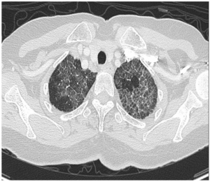
Case 2: CT Scan of patient (b) showing ground glass and interstitial thickening typical of Pulmonary Alveolar Proteinosis
50 year old female had progressive shortness of breath for a year with multiple admissions to various hospitals. Her various episodes were erroneously labelled as pneumonia or congestive heart failure. However, her Chest Xrays (a) never improved from such episodes and her CT Scan is shown (b).
Pulmonary alveolar proteinosis is a rare lung disorder characterised by abnormal surfactant accumulation in the lung alveoli. The accumulated surfactant results in impairment in gas exchange and respiratory failure.
https://rarediseases.org/rare-diseases/pulmonary-alveolar-proteinosis/
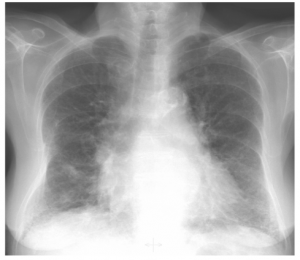
Case 3: CXR (a) showing small lung volumes and bilateral lower lobe predominant lung infiltrates
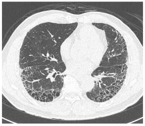
Case 3: CT Scan (b) showing typical basal and peripheral cystic changes of Idiopathic Pulmonary Fibrosis
67 year old Male ex-smoker presents with dry cough and progressive shortness of breath. He was diagnosed as having chronic obstructive pulmonary disease (COPD) or congestive heart failure but did not improve with standard treatment. Instead, he continued to worsen and subsequently needed long term oxygen therapy.
Idiopathic Pulmonary Fibrosis (IPF) is a relatively uncommon disorder which typically afflicts elderly males and ex-smokers. In this condition the lungs become stiffer with permanent scarring. Diagnosis is often difficult with no reliable biomarker and most patients are unfit for any invasive procedures anyway. At this time, there is no cure for this condition.
https://www.nhlbi.nih.gov/health-topics/idiopathic-pulmonary-fibrosis
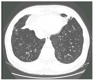
Case 4: CT Scan shows bilateral diffuse bronchiectasis and bronchiolectasis, typical of Diffuse Pan Bronchiolitis
50 year old Male gave a 1 year history of cough, sputum, wheezing and runny nose. He was seen at various clinics and treated as for asthma but never improved with standard therapy.
Diffuse Pan Bronchiolitis is a rare condition first described by the Japanese but has since been described in Asians. They typically present with chronic sinusitis, breathlessness, wheezing and productive cough. Biopsy is often unreliable, but it is important to diagnose it as treatment with long term macrolide antibiotics result in very marked improvement.
https://rarediseases.info.nih.gov/diseases/8526/diffuse-panbronchiolitis
Lung specialist A/Prof Philip Eng, who practises at Philip Eng Respiratory & Medical Clinic, specialises in respiratory and critical care management with a focus on evidence-based medicine and patient care. If you suspect you have a respiratory condition, get in touch with the clinic for more information or to book an appointment.